Vitamin D is obtained from sun exposure, diet (oily fish or fortified dairy products) and dietary supplements. Serum concentration of 25-hydroxyvitamin D [25(OH)D] is a valid marker of vitamin D status.1 Very low levels of 25(OH)D (e.g.
Vitamin D is obtained from sun exposure, diet (oily fish or fortified dairy products) and dietary supplements. Serum concentration of 25-hydroxyvitamin D [25(OH)D] is a valid marker of vitamin D status.1 Very low levels of 25(OH)D (e.g. <20–25nmol/l) have long been recognised as the cause of rickets in childhood and in adults can give rise to skeletal and muscular abnormalities.2 Research in recent years has indicated that vitamin D concentrations not low enough to result in skeletal abnormalities are nevertheless associated with a number of pathological conditions.3 It has therefore been suggested that serum 25(OH)D concentration should preferably be above 75nmol/l.2,4
With this background, hypovitaminosis D may be considered a major health problem, with more than one billion people worldwide having either vitamin D deficiency or insufficiency.2 During recent years, a considerable body of evidence has emerged suggesting that vitamin D may also have an impact on the development of type 2 diabetes (see Figure 1).5–7 Data from the third National Health and Nutrition Examination Survey (NHANES III) revealed that vitamin D deficiency was associated with an increased risk of type 2 diabetes.6 Conversely, in the Nurses’ Health Study, Pittas et al. reported a 33% decreased risk of type 2 diabetes in women with high vitamin D intake compared to women with low intake.8
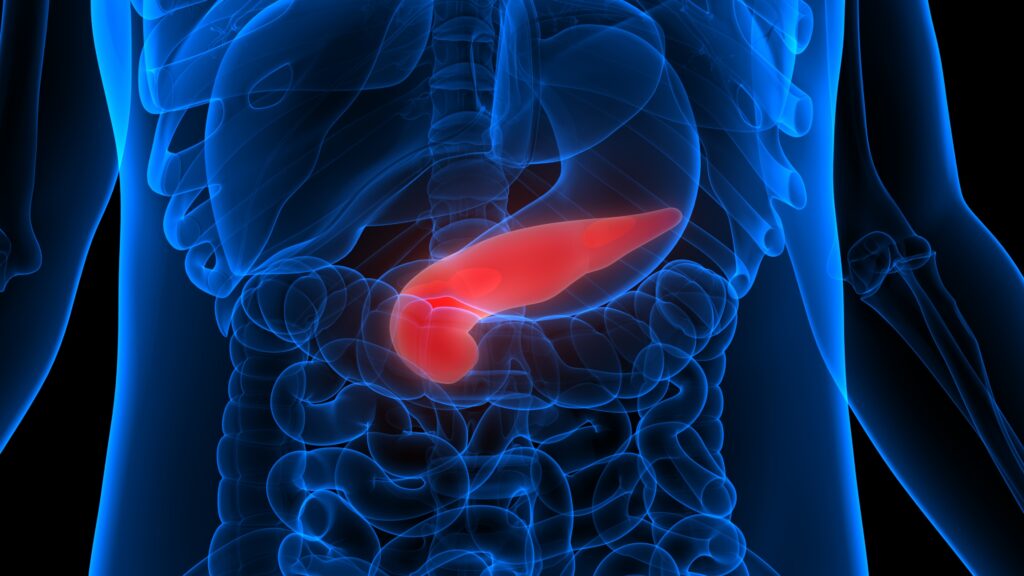
Resistance to the metabolic actions of insulin in the liver and muscle, and insulin secretory dysfunction in the β-cells of the pancreas are the main pathophysiological disturbances that lead to type 2 diabetes. Several other tissues and organs also play important roles in the pathogenesis of the disease, among which fat tissue, the gut with its incretin hormones, the pancreatic α-cells, kidneys and brain may be the most important.9
The exact mechanisms responsible for impaired insulin secretion and action remain to be fully elucidated. In contrast to the situation in type 1 diabetes, where the gradual and usually rapid reduction in insulin secretion parallels a reduction in β-cell mass, there are plenty of β-cells present even after many years of type 2 diabetes.
A major cause of the impaired insulin secretion in type 2 diabetes, therefore, seems to be impairment in glucose-induced insulin secretion, while the response to other secretagogues is better preserved. Accumulation of lipids in the β-cells and increased circulating levels of non-esterified fatty acids (lipotoxicity) and glucose (glucotoxicity) may contribute to impaired insulin secretion.10
Insulin resistance in type 2 diabetes and prediabetes is mainly due to a post-receptor defect in insulin signalling that reduces non-oxidative glucose metabolism. It seems to be associated with mitochondrial dysfunction and/or endoplasmatic reticulum stress and, at least in some tissues, the accumulation of lipid droplets.10
This article explores the possible role of vitamin D deficiency in the pathogenesis of type 2 diabetes. It reviews the literature investigating a potential role for vitamin D in the regulation of insulin secretion and action in subjects with and without type 2 diabetes.
Measuring Insulin Secretion and Action
In the studies reviewed here, measurements of insulin action and secretion have been performed using a variety of different methods. The preferred methods are direct measurements, such as the euglycaemic, hyperinsulinaemic glucose clamp for measuring insulin sensitivity. The hyperglycaemic clamp or intravenous glucose tolerance test (IVGTT), with the use of Bergman’s minimal model, can estimate both insulin sensitivity and secretion. However, these tests are cumbersome and expensive to perform.
In most of the studies referred to herein, more easily measured markers of insulin resistance and secretion have been used. These are usually based on measurements in fasting blood samples and include (among others) fasting insulin, fasting C-peptide and indices that combine fasting measurements of glucose and insulin, such as the HOMA (homeostatic model assessment) and QUICKI (quantitative insulin sensitivity check index) indices. In some instances, indices based on oral glucose tolerance tests (OGTT) have also been used. These include the 30-minute value of insulin, Matsuda index and the oral glucose insulin sensitivity index.
All of these methods have their benefits and disadvantages, and are discussed in further detail elsewhere.11–13
Insulin Action
Several studies describe an association between vitamin D status and insulin sensitivity (see Tables 1 and 2). These are mostly cross-sectional studies that have shown a positive association between serum 25(OH)D concentration and fasting measures of insulin sensitivity,6,14,15 but the results are ambiguous.16,17
By contrast, Manco et al. found no relationship between vitamin D status and insulin sensitivity, measured with the euglycaemic, hyperinsulinaemic clamp, in 116 morbidly obese subjects.20 Likewise, the serum 25(OH)D concentration was not associated with euglycaemic, hyperinsulinaemic clamp-estimated insulin sensitivity in 39 non-diabetic Italians.21 No association with serum 25(OH)D concentration was seen when measuring insulin sensitivity using IVGTT and Bergman’s minimal model in 446 subjects with the metabolic syndrome.22
Kayaniyil and associates investigated the cross-sectional associations between vitamin D and OGTT-measured insulin sensitivity. They found that low levels of serum 25(OH)D were associated with low Matsuda insulin sensitivity index and increased HOMA-insulin resistance (IR).23 Interestingly, in sub-analyses according to BMI, these associations were only valid in those with a BMI <30kg/m2. The influence of BMI could possibly partly explain the differences observed between studies.
The importance of serum 25(OH)D concentration for insulin action in subjects with type 2 diabetes is not clear. In 34 subjects with type 2 diabetes, Orwoll did not find any association of vitamin D status with concentrations of glucose, C-peptide and insulin, whether levels were measured when fasting or meal stimulated.24 Sufficiently large studies using direct measures are, however, lacking.
The effect of vitamin D on insulin sensitivity might be dependent on ethnicity. In NHANES III, serum 25(OH)D concentration correlated negatively with HOMA-IR in Caucasians and Mexican-Americans but not in Afro-Americans.6 In another study, vitamin D intake was positively associated with IVGTT-measured insulin sensitivity and was inversely associated with HOMA-IR in Afro-American women. The relationships were independent of age, total body fat, energy intake and percentage of kcals from fat. No such associations were seen in European- Americans. The study did not report measurements of serum 25(OH)D.25
There are only a few prospective studies on the predictive values of 25(OH)D on glucose metabolism. In a longitudinal cohort study of 524 non-diabetic men and women aged 40–69 years, Forouhi et al. demonstrated an inverse correlation between baseline serum concentration of 25(OH)D and future glycaemia and insulin resistance, measured by HOMA-IR.26 After 17 years of follow-up of the Mini-Finland Health Survey, a relative risk of 0.6 for developing type 2 diabetes was found for the highest, compared to the lowest, 25(OH)D quartile. This association was attenuated, however, after adjustment for BMI and physical activity.7
Intervention with vitamin D supplementation may affect insulin sensitivity (see Table 2). Pittas and co-workers reported on a randomised controlled trial in 314 non-diabetic subjects given cholecalciferol and calcium supplementation or placebo for three years.27 In a subgroup of subjects with impaired fasting glucose, vitamin D supplementation attenuated the increases in glycaemia and insulin resistance measured by HOMA-IR seen in the placebo group. No effect was seen in subjects with normal fasting glucose concentration.27 This was, however, a post-hoc analysis of a trial designed for the prevention of osteoporosis, rather than to determine the effects of vitamin D on insulin sensitivity.
In line with these results is the SURAYA study, where obese, insulin-resistant South-Asian women living in New Zealand were given 4,000IU of cholecalciferol or placebo daily for six months. HOMA-IR was significantly improved, but only in subjects who reached a 25(OH)D serum concentration of >80nmol/l and only after six months of supplementation.28 This may suggest a time- and dose-dependent effect of vitamin D supplementation. This is also supported by the notion that in studies on both bone and muscle, it takes many months of adequate vitamin D supplementation to normalise vitamin D at the tissue level.29
Nagpal and co-workers observed no effect of vitamin D levels on HOMA-IR. They did, however, find a significant effect on three-hour oral glucose insulin sensitivity testing, after an intervention with 120,000IU cholecalciferol given fortnightly for six weeks in obese Asian-Indian men.30
Data on vitamin D supplementation in subjects with type 2 diabetes are scarce and most studies are small. A Scottish, randomised, controlled trial in 34 subjects, given 100,000IU ergocalciferol as a single dose, revealed no effect on HbA1c or HOMA-IR after eight weeks. Participants did, however, have significantly improved flow-mediated dilation.31
Likewise, Jorde and Figenschau did not find an effect on HbA1c or HOMA-IR when giving patients cholecalciferol 40,000IU/week during a six-month placebo-controlled study. This study included subjects with a mean 25(OH)D level of 60nmol/l, however, and had limited power.32
Parekh et al. and Patel et al. observed no effect on insulin sensitivity, QUICKI and HOMA-IR of vitamin D supplementation in 28 Asian-Indian and 24 American subjects with type 2 diabetes, respectively.33,34 Very few studies have used direct measures of insulin sensitivity. Orwoll found no effect on a meal challenge after calcitriol supplementation.24 IVGTT-measured insulin sensitivity did not change in a Danish study of 1-α-hydroxycholecalciferol supplementation in subjects with newly diagnosed type 2 diabetes or impaired fasting glucose.35
Insulin Secretion
Animal and in vitro studies suggest a relationship between vitamin D and pancreatic β-cell function. Rabbits, rats and mice with vitamin D deficiency display impaired insulin secretion that improves with vitamin D supplementation.36–39
The effects on insulin secretion in man, however, are not clear. Anecdotal case reports in the 1980s indicated favourable consequences of vitamin D repletion in subjects with type 2 diabetes.40,41 Despite this, data from cross-sectional (see Table 3) and interventional (see Table 4), studies are sparse and the results are not conclusive.
In a relatively small study among East London Asians with severe vitamin D deficiency, serum vitamin 25(OH)D concentration correlated with insulin and C-peptide concentrations 30 minutes after an OGTT.42 Insulin was not measured at baseline and two hours after glucose intake in this study. For this reason, the 0 to 30 minute increase or the area under the curve have not been calculated. The data were not adjusted for BMI and other covariates either.
Interestingly, when OGTTs were performed in a group of elderly Dutchmen, 25(OH)D concentration correlated inversely, in adjusted analyses, with the area under the curve for glucose. They also correlated inversely for insulin after exclusion of eight subjects with newly-diagnosed type 2 diabetes.14 These results do not suggest an insulin secretory defect in subjects with hypovitaminosis D.
In a cross-sectional study of young, healthy, glucose-tolerant students, Chiu et al. observed an independent negative relationship between serum 25(OH)D concentration and plasma glucose concentration after an OGTT. They interpreted these findings as a β-cell dysfunction.18 The authors then went on to use the more invasive hyperglycaemic clamp method in the same subjects. Here, the initially-observed inverse relationship between both first- and second-phase insulin secretion and serum 25(OH)D concentration was not significant after adjusting for covariates, including BMI.18
In accordance with Chiu et al., Gulseth et al. observed no association between serum 25(OH)D concentration and insulin secretion estimated from an IVGTT as the acute insulin response to glucose or the disposition index. The study included a large sample of European subjects with the metabolic syndrome. No association was seen between vitamin D status and HOMA-β after appropriate adjustment for BMI and other covariates.22
In the large NHANES III study, with more than 6,000 participants, there was no relationship between HOMA-β and serum 25(OH)D concentration either.6 By contrast, Kayaniyil et al. recently reported a statistically significant association between 25(OH)D and insulin secretion measured during an OGTT, although the clinical significance is questionable, based on the given regression equations.23 Possible associations between serum 25(OH)D concentration and measures of insulin secretion have scarcely been investigated in subjects with diagnosed type 2 diabetes. Orwoll et al. reported no relationship between vitamin D status and glucose, C-peptide concentrations and insulin after a meal challenge in 35 subjects with type 2 diabetes.24
In general, measures based on OGTT tend to be positively associated with serum 25(OH)D concentration.14,18,23,42 Fasting measurements (HOMA-β) and more invasive methods, such as the IVGTT and hyperglycaemic clamp, do not show such an association.6,18,22 One can only speculate about the possible relationship between the incretins, insulin secretion and vitamin D levels.43
Data on insulin secretion from vitamin D intervention studies are sparse (see Table 4). Most studies are small and inadequately powered or designed, and use surrogate measures of insulin secretion. In the study by Boucher et al.,42 22 subjects with severe vitamin D deficiency (25(OH)D <27.5nmol/l) were supplemented with cholecalciferol 100,000IU. After eight to 12 weeks both C-peptide and insulin secretion, measured 30 minutes after OGTT, were improved. Borrisova and co-workers reported on an intervention in 10 female subjects with type 2 diabetes given 1,300IU cholecalciferol per day for four weeks. Apparently, first-phase insulin secretion, measured during an IVGTT, was increased after the intervention.44 These studies were not randomised, controlled trials, however.
By contrast, data from currently available randomised, controlled trials do not show an effect of vitamin D on insulin secretion. Neither Lind et al.45 nor Ljunghall et al.35 found any effect of calcitriol intervention on IVGTT-measured insulin secretion in subjects with glucose intolerance. Jorde and Figenschau report of no effect on HOMA-β after six months of cholecalciferol intervention in 32 Norwegian subjects with type 2 diabetes.32 Likewise, cholecalciferol treatment did not influence HOMA-β significantly in insulin-resistant or glucose-intolerant subjects in the SURAYA trial from New Zealand and a trial from New Delhi, India.28,30
Possible Mechanisms Linking Vitamin D to Insulin Action and Secretion
Even though the clinical evidence of vitamin D effects on insulin secretion and action are indecisive, there are biological mechanisms that could explain such an effect. Many tissues, including the pancreas, express vitamin D receptors and also have the ability to convert 25(OH)D to its active form 1,25(OH)2D by the expression of 25-hydroxyvitamin D-1-α-hydroxylase.3
As intracellular calcium concentrations and calcium fluxes over the cellular membrane are important regulators of insulin secretion, it has been suggested that vitamin D may exert its effects on the β-cell by its ability to regulate calcium. Calcium is also essential for insulin action in target tissues, so the association of insulin resistance with low vitamin D-levels may be due to impaired transduction of the intracellular signalling pathway after insulin binds to its cellular receptor. Alternatively, inflammation associated with accumulation of intra-abdominal fat seems to be an important mediator of both insulin secretory dysfunction and insulin resistance.10 The major role of vitamin D on immune cells may therefore be key to its antidiabetic effects.
Genetics
The vitamin D receptor acts as a transcription factor when bound to 1,25(OH)2D. Polymorphisms in the vitamin D receptor gene – TaqI, ApaI, FokI and BsmI – may influence insulin action and secretion, although data are sparse and inconclusive.46
In Bangladeshi Asians the TaqI polymorphism has been associated with insulin secretion,47 whereas the BsmI gene variant was related to insulin resistance measured as the HOMA-IR index in Caucasian- Americans.48 In general, no statistical differences in vitamin D receptor gene polymorphism frequencies have been found between subjects with type 2 diabetes and controls.49–51 Data using direct measures of insulin sensitivity and secretion and investigations comparing different ethnicities are currently lacking. The active vitamin D metabolite, vitamin 1,25(OH)2D, circulates bound to its specific vitamin D binding protein. Genetic variants of this binding protein have been linked to an increased risk of developing type 2 diabetes and/or insulin resistance in several,52–54 but not all,52,53 populations. Polymorphisms in vitamin D-related genes could possibly explain the observed differences between populations, both in response to vitamin D supplementation and the cross-sectional associations between serum 25(OH)D concentration and insulin action and secretion.
Conclusions
In conclusion there is some, but not definitive, evidence that low levels of vitamin D may be causally related to insulin resistance. The evidence that links hypovitaminosis D to insulin secretory dysfunction seems to be weak and mostly indirect. There is a need for prospective, clinical, intervention studies applying up-to-date direct measurements of insulin action and secretion in subjects with low levels of vitamin D to clarify the issue.


