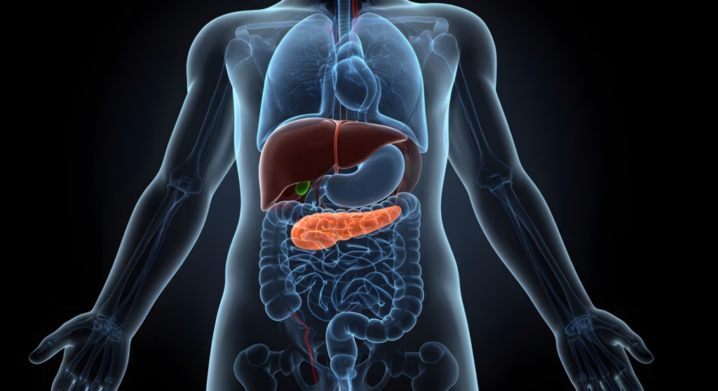
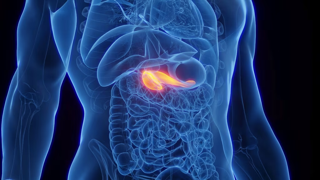
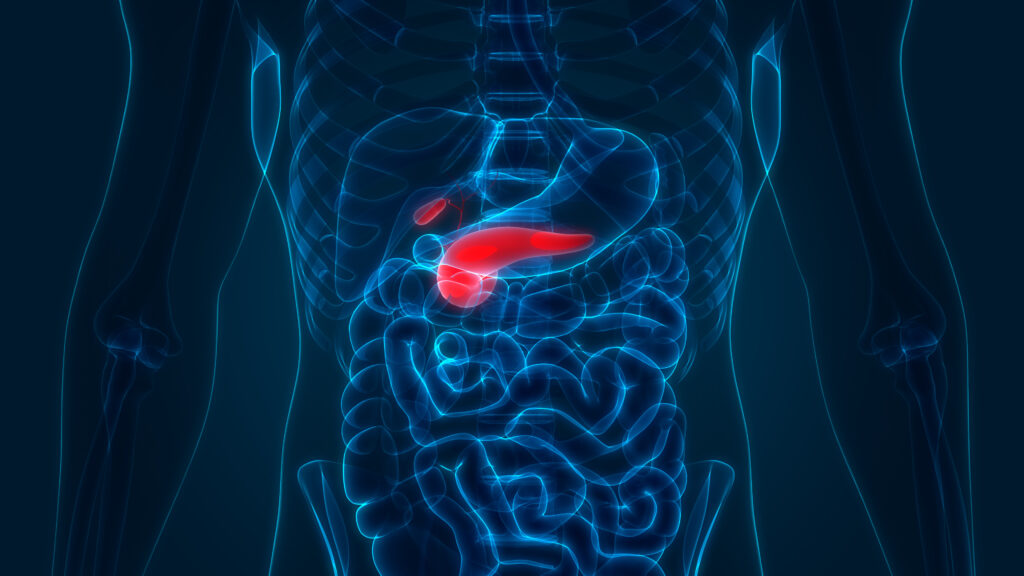
According to the last American Diabetes Association (ADA) recommendations, it is possible to demonstrate an abnormality in carbohydrate metabolism, diagnostic for diabetes, by measurement of fasting plasma glucose (glycaemia ≥126mg/dl, i.e. ≥7.0mmol/l) or after a challenge with an oral glucose load (OGTT) (glycemia ≥200mg/dl two hours post an oral 75g glucose challenge, i.e. ≥11.1mmol/l) or symptoms of diabetes plus casual plasma glucose concentration ≥200mg/dl (≥11.1mmol/l).
According to the last American Diabetes Association (ADA) recommendations, it is possible to demonstrate an abnormality in carbohydrate metabolism, diagnostic for diabetes, by measurement of fasting plasma glucose (glycaemia ≥126mg/dl, i.e. ≥7.0mmol/l) or after a challenge with an oral glucose load (OGTT) (glycemia ≥200mg/dl two hours post an oral 75g glucose challenge, i.e. ≥11.1mmol/l) or symptoms of diabetes plus casual plasma glucose concentration ≥200mg/dl (≥11.1mmol/l). In the absence of unequivocal hyperglycaemia, these criteria should be confirmed by a second measurement of blood glucose levels obtained on a different day.1 These glucose diagnostic levels are the results of a re-examination of the classification and diagnostic criteria of diabetes made in 1997 by an international expert committee on the basis of the 1979 publication of the National Diabetes Data Group and the subsequent World Health Organization (WHO) study group. In 1979, in fact, the US Diabetes Data Group (also known as the NDDG) introduced innovations into diagnostic criteria for diabetes based for the first time upon epidemiological studies that stated a correlation between blood glucose values and burden of diabetic complications such as retinopathy and nephropathy.2 Diagnostic blood glucose level was fixed at 140mg/dl (7.78mmol/l); if glycaemia was <140mg/dl but >110mg/dl, an OGTT was required (diagnostic if glycaemia >200mg/dl two hours post-OGTT). In 1980 these criteria were also adopted by the WHO, with small variations.3 In 1997, an expert committee of the ADA further revised diagnostic criteria for diabetes: the cut-off of fasting plasma glucose (FPG) was lowered from 140mg/dl (7.78mmol/l) to ≥126mg/dl (≥7.0mmol/l) on the basis of population studies showing that the incidence of rethinopathy was significantly higher even with blood glucose levels ≥120mg/dl.4
These criteria were recently confirmed in 19995 and in 20036 by the expert committee of ADA; this new cut-off of FPG reduces the discrepancy between fasting and two hours post-OGTT blood glucose levels for diabetes diagnosis, promoting the preferential use of FPG in place of OGTT for diagnostic purposes, since OGTT is burdened with greater costs, less reproducibility and technical difficulties. Referring to the above-mentioned studies, the chosen FPG value was shown to have the same predictive power of two hours post-OGTT blood glucose. On the other hand, the European DECODE study (Diabetes Epidemiology: Collaborative Analysis of Diagnostic Criteria in Europe) shows that two hours post-OGTT blood glucose is morepredictive than FPG in relation to cardiovascular disease.7In fact, evidence that allows us to say which test is better for diagnosing diabetes is not available; therefore, according to the ADA expert committee, FPG remains the test of choice in clinical practice, while OGTT might be useful in research studies or in clinical situations that require a clear distinction between diabetes and other glucose metabolism as defined below. Besides a new threshold for FPG, ADA identified a new diagnostic category – impaired fasting glucose (IFG) – for fasting blood glucose levels between 110 and 125mg/dl. This category is different from impaired glucose tolerance (IGT), already defined in previous criteria, and confirmed as blood glucose levels between 140 and 199mg/dl two hours post-OGTT. These two blood glucose abnormalities do not indicate two defined clinical conditions, but they represent a risk of a possible evolution towards diabetes itself and, apart from diabetes, a risk of cardiovascular complications. Nowadays it is matter of debate whether to change all of these cut-offs, lowering either FPG or the limit for IGT. The choice of a lower cut-off could be made considering not only epidemiological data that seem to show that the limit of 110mg/dl is too high for IFG in order to predict future diabetes onset, but also other factors, such as the ratio of benefits to costs in an individual who undergoes the risk of diabetes. For these reasons, ADA actually suggests that IFG cut-off point should be reduced from 110 to 100mg/dl, while the IGT cut-off point should not change.
In the last few years there has been some discussion on the possible role of glycated haemoglobin (HbA1c) as a diagnostic test for diabetes. HbA1c represents an average of blood glucose levels in a lag time of three months, so when it is elevated it is able to indicate a chronic state of high blood glucose levels. Moreover, as for FPG and for two hours post-OGTT, there is also a threshold level of HbA1c associated with risk of retinopathy.3 Otherwise, there are some disadvantages – such as, for example, different clinical conditions that may influence HbA1c, different assay methods and different reference ranges – so ADA does not recommend HbA1c as a diagnostic test for diabetes, even if it is useful to check the efficacy of glucose-lowering therapy.
Classification of Diabetes Mellitus
When abnormal blood glucose levels have been identified as diabetes, it is important to categorise the type of diabetes for clinical and therapeutical purposes. The most recent ADA recommendations have tried to make a classification based, whenever possible, on aetiological features. Certainly, the two main categories are represented by type 1 diabetes (formerly known as insulin-dependent diabetes mellitus, IDDM) and type 2 diabetes (formerly known as non-insulin-dependent diabetes mellitus, NIDDM).Type 1 diabetes recognises an immunological pathogenesis with progressive β-cell destruction that leads to little or absent insulin secretion. The lack of insulin may lead to the presence of ketones in blood and urine (before insulin therapy is started) and may be confirmed by a glucagon test. A blood sample, in fasting condition and with a blood glucose level <200mg/dl, is taken; then, after the infusion of 1mg intravenous (IV) glucagon, a second blood sample is taken after six minutes. Post-glucagon C-peptide levels <0.6nmol/l are considered indicative of insulin deficiency.8 Autoimmune β-cell damage may be measured by the presence of antibodies such as islet cell autoantibodies (anti-ICA), autoantibodies to glutamic acid decarboxylase (anti-GADA), autoantibodies to protein tyrosine phosphatase isoforms (anti- IA2) or autoantibodies to insulin (anti-IAA).
Type 2 diabetes, the most frequent form of diabetes, is due to insulin resistance together with a relative insulin deficiency. It is often associated with overweight and obesity and until a few years ago it occurred mainly in adults; now, its appearance in young age is rapidly increasing due to the high prevalence of obesity at this age as well. A significant proportion of type 2 diabetic patients remain undiagnosed for years; since the earlier the diagnosis is made, the easier it is to prevent diabetes complications, it is recommended, according to ADA, to periodically screen non-diabetic individuals ≥45 years of age, particularly those who are overweight (body mass index (BMI) > 25kg/m2). Screening has to be performed especially with FPG and only in some cases with OGTT. If negative, the screening has to be repeated every three years.
The screening has to be anticipated in younger people or performed more frequently in people who are at high risk of developing diabetes, such as: overweight people (BMI >25kg/m2); first-degree relatives of type 2 diabetic patients; women with gestational diabetes mellitus (GDM –see below) or with macrosomic foetus (>4kg); people with hypertension, high-density lipoprotein (HDL) cholesterol <35mg/dl and/or triglycerides >250mg/dl; previous IFG or IGT; clinical conditions associated to insulin resistance (i.e., acanthosis nigricans); people with vascular diseases; or particular ethnic groups (i.e. Afro-Americans).
Between type 1 and type 2 diabetes, there is a condition, often unrecognised, in which some features of these principal categories are mixed – latent autoimmune diabetes of the adult (LADA). LADA is a type of diabetes occurring in adults in whom autoimmune β-cell damage evolution is slower than in children, so they could be diagnosed as ‘type 2’ by mistake, since at the beginning of disease there is not clear insulin deficiency. These diagnostic problems have some prognostic and treatment implications as advised by the UK Prospective Diabetes Study (UKPDS),9 because patients with LADA are prone to insulin deficiency and may need insulin therapy earlier than patients without LADA. When LADA is suspected, diagnosis may be confirmed by the presence of autoantibodies positive to GADA, together with IA2A or ICA.Another form of diabetes is the maturity onset diabetes of the young (MODY). This is a form of diabetes associated with several monogenetic defects that are inherited as autosomal dominant pattern; these abnormalities induce defects in insulin secretion. This form of diabetes may be suspected by the presence of a family history, without generational jump, for at least three generations, and by onset before 25 years of age.
Among the different types of diabetes, GDM is very important from a clinical point of view. According to the ADA position statement, GDM is: “any degree of glucose intolerance with onset or first recognition during pregnancy”. The definition applies regardless of whether insulin or diet-only therapy is used for treatment, or whether the condition persists after pregnancy. Since GDM is associated with a high risk of complications (macrosomal foetus, hypoglycaemia, respiratory distress syndrome, polyidramnios, etc.), it is necessary to screen for this condition. Screening for GDM should be performed in all women except those who are at low risk, as per the following characteristics: <25 years of age; normal body weight; no family history of diabetes; no history of poor obstetric outcome; or not member of an ethnic/racial group with a high prevalence of diabetes. Screening for GDM should be performed at the first prenatal visit for women at high risk (marked obesity, personal history of GDM, glycosuria or a strong family history of diabetes) and should be repeated, if necessary, at 24–28 weeks of gestation; in women with average risk, screening should be performed only at 24–28 weeks of gestation. The first test to perform is FPG, or casual plasma blood glucose levels: if the results of these tests do not meet diagnostic criteria for diabetes, and in the absence of unequivocal symptoms, a second test is required. It is possible to perform two types of test: a 50g oral glucose load, followed, if positive, by a 100g oral glucose load, or a 100g oral glucose load only. When the two-step approach is used, a glucose threshold value >140 mg/dl after one hour of a 50g load identifies about 80% of women with GDM; the sensitivity of this test rises to 90% when the cut-off is lowered to 130mg/dl. Diagnostic criteria for 100g oral glucose load are: 95mg/dl at fasting, 180mg/dl after one hour, 155mg/dl after two hours and 140mg/dl after three hours; the test is considered positive if at least two values are higher or equal to these thresholds.1 Instead of a 100g oral glucose load test, it is possible to perform a 75g oral glucose load test, although this test has been less validated in pregnancy. Diagnostic levels for the 75g oral glucose load are: 95mg/dl at fasting, 180mg/dl after one hour and 155 mg/dl after two hours. After pregnancy, women with GDM should be reclassified. In fact, they could continue to have diabetes, they could return to normal glucose tolerance or they could continue to have IFG or IGT.There are many other types of diabetes in which diagnosis is made by the presence of other conditions, including: diabetes secondary to pancreatic diseases (such as chronic pancreatitis, haemochromatosis); endocrine diseases with an excess of counterregolatory hormones (such as Cushing’s syndrome, acromegaly, glucagonoma, pheochromocytoma); use of diabetogenic drugs (such as glucocorticoids, pentamidine, Vacor, diazoxide, thiazide); virus infections (such as congenital rubella, cytomegalovirus); some genetic syndromes (such as Down, Klinefelter, Turner, Prader-Willi); and particular conditions of insulin resistance (such as leprecaunism, insulin resistance type A).


