Since the description of Turner syndrome (TS) in 1938, a wealth of information has been added and our current understanding of the syndrome is continuously being broadened. The syndrome affects only females and care must include the close collaboration of several specialties such as genetics, embryology, paediatrics, gynaecology and obstetrics, endocrinology, cardiology, gastroenterology, otorhinonology and ophthalmology.
Since the description of Turner syndrome (TS) in 1938, a wealth of information has been added and our current understanding of the syndrome is continuously being broadened. The syndrome affects only females and care must include the close collaboration of several specialties such as genetics, embryology, paediatrics, gynaecology and obstetrics, endocrinology, cardiology, gastroenterology, otorhinonology and ophthalmology.
In this article, the focus is on the diverse clinical aspects, including epidemiology, endocrinology, cardiology, gastroenterology and gynaecology of the syndrome, with reference to recent genetic discoveries.
Diagnosis, Genetics and Epidemiology
There are no firm guidelines for the diagnosis of TS. However, the generally accepted cardinal stigmata include growth retardation with reduced adult height and, except in rare cases, gonadal insufficiency and infertility. The genetic background of the TS phenotype is highly variable, including complete or partial absence of the sex chromosomes (the X and/or Y chromosomes). In addition to this, mosaicism with two or more cell lines may be present. The first described cases were of the ‘classic’ karyotype 45,X. In more recent series the classic karyotype has accounted for 50% of cases; the remaining cases comprise mosaic karyotypes, karyotypes with an isochromosome of X – for example i(Xq) or i(Xp) – or karyotypes with an entire or part of a Y chromosome.1 The genetic basis for the traits of TS is continuously being unravelled as the functions of the hort stature homeobox (SHOX) gene become clearer. Haploinsufficiency of SHOX explains the reduction in final height, changes in bone morphology, sensorineural deafness and other features. Additional genes are thought to be involved in the pathogenesis of TS, but await discovery.
Prenatal prevalence of the syndrome is much higher than post-natal prevalence, owing to a well-described increased intrauterine mortality. Prenatal diagnosis of TS may not always be correct. Therefore, a more precise diagnosis depends on the inclusion of high-resolution ultrasound scan, foetal echocardiography and other advanced modalities. A European multicentre study found an induced abortion rate of 66%, and thus most diagnosed foetuses with TS are legally aborted. This study confirms previous studies showing legal abortion rates of 60–80%.1 However, this is only a fraction of foetuses with TS since less than 10% of any pregnant population is subjected to invasive methods of prenatal diagnosis, and the use of prenatal ultrasound scanning will lead to diagnosis of only the cases with the most pronounced phenotype, i.e. hydrops or increased nuchal fold.1 Figures for the prevalence of TS are based on a number of cytogenetic studies, with estimates ranging from 25 to 210 per 100,000 females,2 giving an estimated proportion of about 50 per 100,000 females in Caucasian populations.
Most post-natal diagnoses are made at birth (15%), during teenage years (26%) or in adulthood (38%), with the remainder of cases being diagnosed during childhood.3 As a result, there is a considerable delay in diagnosing girls and adolescents (see Figure 1).4 The key to diagnosis is lymphoedema in 97% during infancy and short stature in 82% in childhood and adolescence.3
Morbidity is considerably increased in TS. In a study of all females diagnosed with TS compared with the general female population, we compared the incidence rates of specific diseases suspected to occur at an increased frequency in TS. The relative risk (RR) of an endocrine diagnosis in TS patients was increased to 4.9 overall: hypothyroidism (RR 5.8), type 1 diabetes (RR 11.6) and type 2 diabetes (RR 4.4). The risk of ischaemic heart disease and arteriosclerosis (RR 2.1), hypertension (RR 2.9) and vascular disease of the brain (RR 2.7) was also increased. Furthermore, the risk of other conditions such as cirrhosis of the liver (RR 5.7), osteoporosis (RR 10.1) and fractures (RR 2.16) was also increased, as was the risk of congenital malformations of the heart, the urinary system and the face, ears and neck. The risk of all cancers was comparable to that seen in the general female population.1 In addition to morbidity, mortality is increased in TS. In a British cohort study, the RR of premature death was increased to 4.25 owing to disease in the nervous, digestive, cardiovascular, respiratory and genito-urinary systems. Death owing to cancer was lower than expected, corroborating the morbidity studies. We found a comparable increase in mortality among Danish patients, and there were important differences between patients with 45,X or an isochomosome, who had a four-fold increase in mortality, and patients with other karyotypes, who had only a two-fold increase in mortality.4
Turner Syndrome and the Heart
Diseases of the heart account for 50% of the excess mortality in TS.6 A number of left-sided cardiovascular malformations such as elongated transverse aortic arch are seen in 50% of TS women,7,8 bicuspid aortic valves are seen in 13–43% compared with 1–2% in the general population9 and 4–14% have aortic coarctation in TS (see Figure 2).1 Less commonly, right-sided malformations and abnormalities such as persistent left vena cava superior and partial anomalous venous return are encountered.7 These congenital heart defects may co-segregate at least to some extent, and in some cases they may severely affect the left ventricle, the outflow tract and the thoracic aorta in hypoplastic left heart syndrome.
Some of these defects call for early and potentially life-saving high-risk cardiovascular intervention, thereby contributing to both increased mortality in infancy as well as later on in life as a result of complications caused by the congenital abnormalities, disease and the necessary interventions.4 Other defects present less acutely, with symptoms associated with late life morbidity. Then again, some women remain subclinical throughout their entire lifetime, with an unresolved place in the pattern of morbidity and mortality in TS. More than 30% of young girls and adolescents and 50% of adults with TS are mildly hypertensive on 24-hour ambulatory measurements. Additionally, 50% have abnormal circadian blood pressure profiles.10–12 Although the pathological nature underlying the hypertensive disorder in TS remains unknown, it is uniformly accepted that hypertension and the sympathovagal dysfunction of nocturnal ‘non-dipping’ confer a high risk of future cardiovascular events, as also epidemiologically documented in TS.4 The presence of these risk factors adds to the other adverse indices of cardiovascular risk in TS: increased carotid intima thickness,13 a propensity towards glucose intolerance and type 2 diabetes,11,14 non-alcoholic steatohepatitis (NASH),15–17 discordant lipid profile,18,19 oestrogen deficiency and unfavourable body composition.20
In addition to the congenital structural cardiac malformations such as bicuspid aortic valve and coarctation of the aorta, hypertension is also thought to be a major factor,21 as shown by the high incidence of aortic dissection and rupture in TS: 40 per 100,000 TS syndrome-years versus six per 100,000 general population years. Strikingly, aortic dissection affects TS patients at a median age of 35 years as opposed to 71 years in the general population (see Figure 3).22 Aortic dilation normally precedes dissection and rupture, and such an abnormal aortic calibre is seen in 3–42% of randomly selected TS women23,24 increasing with age.25 Aortic calibre correlates with systolic blood pressure, but surprisingly, not with vascular atherosclerotic indices such as aortic stiffness or plasma lipids21 in TS. A hitherto unknown intrinsic arterial defect is highly likely to be part of the generalised vasculopathy in TS.13,26
Subclinical systolic and severely impaired diastolic function associated with increased left atrial dimensions is seen even in normotensive and strictly metabolically controlled TS women.27 Furthermore, N-terminal pro-brain natriuretic protein (BNP) is elevated in the absence of symptoms of cardiac failure and no systolic dysfunction,12 which is interesting in light of the predictive capacity of this cardiac neurohormone in heart failure. An intrinsic cardiac dysfunction is supported by the prolongation of the QTc interval seen in 30% of girls, adolescents and adults with TS,28 a condition accepted as an independent predictor of sudden cardiovascular death.
Premature ovarian failure (POF) is the most prevalent cardiovascular risk factor in TS. It is currently unclear precisely how early oestrogen deficiency has an impact on cardiovascular prognosis, although young age at menopause in other populations confers a more adverse risk profile for events. Evidence confirming earlier observational findings of oestrogen-derived cardioprotection is supported by animal and human studies showing anti-inflammatory, antioxidant and lipid-lowering effects with modification of disruptive vascular processes. On the other hand, balanced against such protective effects is the potential for the induction of myocardial hypertrophy, venous thromboembolic events and proarrhythmic territories.29 In interventional studies, oral hormone replacement therepy (HRT) did not reduce the total risk of cardiovascular disease in primary and secondary prophylaxis of atherosclerosis in post-menopausal women, while the potential alleviation of adverse risk in POF has not been investigated. However, evidence for alleviation of cardiovascular disease risk with the early introduction of HRT in those with deficiency of female sex steroids early in life is mounting – the so-called ‘timing hypothesis’.29
The congenital and acquired cardiovascular morbidities occur against a backdrop of endocrine problems, creating highly variable cardiac phenotypes where neither risk prediction nor modification is straightforward. Therefore, all girls and women with TS should be regarded as facing an increased risk of cardiovascular events,30 and it is recommended that TS girls and women be submitted to a baseline delineation of cardiac phenotype and regular follow-ups by cardiologists trained in congenital heart disease.31 Depending on the presentation, examination should include an electrocardiogram, 24-hour ambulatory blood pressure, echocardiography and magnetic resonance imaging.31
Ovarian Insufficiency and Hormone Replacement Therapy
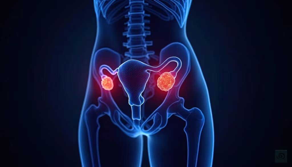
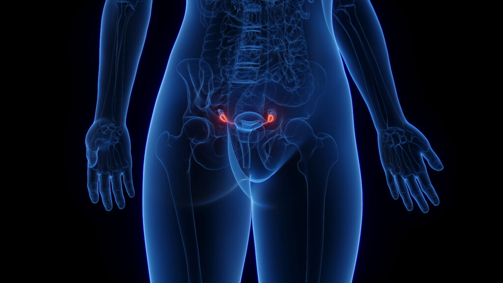
TS belongs to a number of conditions that are collectively termed ‘premature ovarian failure’. Early ovarian demise presents in most patients with TS, with ensuing oestrogen insufficiency. The ovarian germ-cell count is normal until week 18 of gestation, after which accelerated degeneration takes place. High levels of follicle-stimulating hormone (FSH) and luteinising hormone (LH) are present in early childhood (two to five years of age) and after the time of normal puberty onset (11 years of age). In adulthood, levels of FSH and LH increase to menopausal levels. Many untreated girls show signs of puberty and/or have regular periods for a varying length of time.32 This may be explained by new data showing that even in some 45,X patients, follicles can still be found in 12–19-year-olds.33 Antimüllerian hormone (AMH) and inhibin B are emerging as possible markers of ovarian function.34,35 Understanding the processes in early follicular apoptosis in TS may in the future lead to a treatment that spares the follicles and maintains fertility. Ideally, the timing of endocrine therapy should allow onset of puberty at the same time as the peers of the patient to avoid social problems secondary to delayed physical and psychological development. This would also allow optimal bone mineralisation to take place. In most normal girls, puberty starts at around 12 years of age. Since 30% of girls with TS undergo some spontaneous pubertal development and 2–5% have spontaneous menses with a small potential to achieve pregnancy without medical intervention.32 signs of puberty should be looked for before starting oestrogen therapy. When FSH and LH are clearly elevated and AMH and inhibin B are low, and where clinical signs of puberty are lacking, pubertal induction should be started, always considering individual circumstances. To induce pubertal development, the dosing and timing of oestrogen therapy should aim at mimicking normal pubertal development. Doses should be individualised, starting with very low doses of oestrogen as monotherapy either orally or percutaneously.36,37 The treatment can be monitored using the development of secondary sex characteristics (Tanner staging), serum LH and FSH, bone maturation or uterine volume. A gestagen is added when breakthrough bleeding occurs. Currently, it is not clear which gestagen is the most advantageous.
Oestrogen therapy should always be co-ordinated with the use of growth hormone (GH). This should be individualised for each patient to optimise both growth and pubertal development. When growth is a priority, delaying oestrogen therapy is an option to avoid compromising adult height. However, recent growth-promoting trials have documented that the physiological timing of oestrogen therapy does not compromise adult height when GH therapy is started early and the dose is increased in a stepwise fashion.38 Proper oestrogen replacement is also pivotal in TS since it has a positive effect on motor speed and verbal and non-verbal memory and processing when administered in puberty. Females with TS present with a particular neurocognitive profile, with impaired performance on motor tasks and impaired visuo-spatial ability but normal verbal skills.39 The deficits in cognition are likely caused by haploinsufficiency of X-linked genes that normally escape X-inactivation, but these putative genes await further elucidation. Infertility is rated as the most prominent problem in adult women with TS.40 Oocyte donation is an option in many countries and fertility preservation is an emerging option. The most recent studies show good results comparable to thse seen with oocyte donation in other infertile patients, although better preparation of the uterus for implantation (uterine size and endometrial thickness) with prolonged treatment with high daily doses of estradiol (4–6mg or up to 8mg of 17β-estradiol) may improve results in TS. Importantly, it is a high-risk endeavor for a TS woman to go through pregnancy, especially in relation to lesions related to the cardiovascular system but also secondary to endocrine features. Androgen insufficiency is present,41 and it could be interesting to evaluate the possible benefits of androgen substitution in TS. During adulthood it is important to continue HRT, even though a number of issues such as dose during the different ages, administration route, type of oestrogen and type of gestagen are unresolved. Female hypogonadism is related to a number of other conditions, and HRT may reduce or completely alleviate their risk. Currently, new studies indicate that the traditional dose of 2mg of estradiol used in TS and other conditions of POF may be too low for normalising the cardiovascular system and for normal growth of the uterus.42,43 Available data on different routes of administration in TS have not demonstrated the advantages of any particular route:11,15,44 newer studies point towards advantages with transdermal or subcutaneous application45–47 but additional studies are needed to fully resolve this issue. A few studies have compared the contraceptive pill with physiological 17β-estradiol and a gestagen, and suggest that contraceptive pills should not be used in the long-term treatment of TS.48,49
Decreased Stature in Turner Syndrome
Short stature is the cardinal finding in girls with TS, affecting 95–99%.50 Growth retardation is already present in utero with birth weight approximately one standard deviation below expected. Furthermore, growth is retarded in infancy and childhood, where height is two standard deviations below normal and, due to the lack of a pubertal growth spurt height at 14 years of age, the heights encountered are approximately four standard deviations below expected if GH treatment is not initiated.51 Overall, the growth phase is prolonged (if puberty is not induced), achieving a spontaneous final height of almost three standard deviations or about 20cm below normal height. Part of the explanation for the small final height relates to the action of the SHOX gene located to the PAR1 region of the X and Y chromosome; haploinsufficiency of the SHOX gene leads to reduced final height, as documented in Leri-Weill dyschondrosteosis. However, the height difference is only about 50–75% of that seen in TS. Therefore, SHOX deficiency can explain only part of the height deficit in TS.52 BNP is a transcriptional target of SHOX and is present alongside SHOX at the growth plate in proliferative chondrocytes.53 Whether the lack of SHOX-induced BNP is involved in cardiovascular malformations and diseases in TS remains to be elucidated.
GH concentrations in TS are found to be normal in some and reduced in others, and it is generally concluded that girls with TS have a growth deficiency with reduced sensitivity to GH rather than a GH deficiency. GH treatment can increase growth velocity and final height. The effect is dose-dependent, and normalisation of final height has been shown to be obtainable with high doses.38 In the only randomised controlled study using GH treatment (0.3mg/kg/week) or placebo, a significant increase in final height of 7.2cm (CI 6.0–8.0) was seen, with HRT started at 13 years of age.54
In cases where the TS diagnosis is made early (before one to two years of age), very early treatment with GH should be instituted. In a randomised, controlled, open-label study including girls with TS nine months to four years of age for GH treatment, it was possible to correct growth failure and promote growth catch-up, bringing 93% of the toddlers back into the normal height range of the background population within two years. By contrast, those in the placebo group were faced with progressive growth failure.55
Besides effects on growth and final height, GH treatment also has beneficial effects on body composition, with reduced fat mass and an increase in lean body mass. Concerns about the effect of GH on the heart have arisen from evidence that left ventricular hypertrophy is found in acromegalic patients with high levels of GH. A Dutch study showed no signs of left ventricular hypertrophy and no increase in blood pressure in TS girls undergoing seven years of GH treatment.56 Gastroenterology and Hepatology
Increased concentrations of liver enzymes, especially alkaline phosphatase, alanine/aspartase aminotransferase and γ-glutamyl transferase (markers of hepatic cell lesion or turnover), are frequent in TS. Bilirubin (excretion) and coagulation parameters (production) are, in most cases, within the normal range.
In a liver biopsy study in 27 women with TS biopsied because of persistently elevated liver tests,57 multiple abnormalities were found, including marked nodular regenerative hyperplasia, multiple focal nodular hyperplasia and cirrhosis, associated in some with obliterative portal venopathy. Other patients showed more moderate changes, including portal fibrosis, inflammatory infiltrates and non-alcoholic fatty liver disease. The authors conclude that the main causes of liver abnormalities in TS are vascular disorders thought to be congenital in origin and non-alcoholic fatty liver disease, without signs of liver toxicity from concomitant oestrogen therapy. This study is important for several reasons: it is the largest; it includes liver biopsies as well as thorough evaluation of other causes of liver disease; it excludes viral, autoimmune and alcoholic causes; and it excludes oestrogen therapy as a player in liver abnormalities. HRT normalises measures of liver function, while dynamic liver tests are normal and not affected by HRT in TS.17 Inflammatory bowel disease (IBD) also seems to be more frequent in TS (2–3%) and should especially be suspected in girls not responding to GH therapy. Coeliac disease is present in 8% of patients58,59 and, since it may cause additional growth stunting, it should always be excluded. The usual guidelines should be followed for IBD and coeliac disease.
Glucose Metabolism and Type 2 Diabetes
Both type 1 and type 2 diabetes occur more frequently in women with TS. Early reports of impaired glucose tolerance (IGT) in TS are found and IGT has been reported in both girls and women with TS. An epidemiological study including 594 TS women found an increased relative risk of both type 1 and type 2 diabetes.1
Generally, fasting glucose levels in TS women are not significantly different from in controls, but fasting hyperinsulinaemia has been documented. During an oral glucose tolerance test (OGTT), IGT has been found in 25–78%11,14 of adult TS patients. In addition to higher glucose levels, the insulin response is increased and some have found a delayed insulin peak during an OGTT. The impaired glucose homeostasis seems to be explained by a decreased insulin sensitivity as well as a reduced ‘first-phase insulin response’, which could be viewed as an inappropriately low β-cell response.11,14 Body composition is distinctly altered in TS, with increased body mass index, decreased muscle mass, increased total fat mass and increased visceral fat mass. A more sedentary lifestyle and decreased VO2max are also found in this population. All factors contribute to the risk of developing reduced insulin sensitivity and diabetes.
Appropriate HRT in TS also seems to be important for glucose homeostasis, even though the findings in TS are not uniform. HRT reduced fasting glucose and fasting insulin60 and, while not improving insulin sensitivity, fat free mass and physical fitness did increase; both latter factors improve glucose homeostasis. By contrast, more subjects were found to have IGT during an OGTT while receiving HRT.11 On balance, HRT may slightly improve glycaemic control.
Insulin levels, both fasting and as an OGTT response, increase during GH treatment. Insulin levels decrease after termination of GH, but remain higher than pre-treatment levels. GH therapy reduces insulin sensitivity, although this effect subsides with cessation of treatment. GH generally reduces insulin sensitivity in the first six to 12 months of treatment, whereafter it stabilises. This stabilisation could be due to changes in body composition with increase in lean body mass and decrease in fat mass. The proportion of TS patients with IGT does not seem to increase significantly during treatment, and glycated haemoglobin (HbA1c) remains unchanged or even decreases during GH therapy. While most of the effects on glucose metabolism seem to reverse after cessation of GH treatment, the long-term effects of the GH-induced hyperinsulinism and insulin resistance are not known.
In the face of widespread abnormalities in glucose homeostasis and an increased risk of type 1 and type 2 diabetes, there is a need to pay attention to the increased risk of impaired glucose homeostasis and diabetes in TS. Recommendations for diagnosis and treatment of diabetes should adhere to general population guidelines. However, yearly screening of fasting glucose should be carried out and, in cases of suspicion, an OGTT should be performed. Bone Diseases
Peak bone mass depends on a number of factors, such as genetic background, nutrition, physical activity, local growth factors and a spectrum of hormones. Estradiol secretion is clearly deficient in childhood and adolescence. Children and younger and middle-aged adult patients with TS have low bone mineral density (BMD) and studies show that fracture risk is increased.1,61,62 pointing towards the major clinical manifestation of the decreased BMD. HRT is considered crucial to avoid a rapid decrease in BMD and to induce maximal peak bone mass in adolescents and young adults.63 This is supported by longitudinal studies of oestrogen-deficient and oestrogen-replete adolescents with TS. Furthermore, patients with spontaneous menstruation have normal BMD, whereas the absenc of menarche is associated with a reduced BMD. A three-year longitudinal study of 21 women with TS (20–40 years of age), with iliac crest biopsies before and three years after treatment with HRT, showed marked effects of oestrogen on bone.
Treatment consisted of estradiol implants (and an oral gestagen cyclically).46 resulting in comparable estradiol levels to those seen in premenopausal women, and considerably higher than levels achieved with regimens used hitherto (estradiol 2mg orally or equivalent transdermal doses). Bone biopsies pointed towards an anabolic effect on the skeleton of estradiol in young TS patients.46 Furthermore, GH may improve BMD. In a recent seven-year study with GH treatment given at three different doses, BMD increased in a dose-dependent manner. However, oestrogen was added after four years of GH treatment, and it is difficult to ascertain the individual effects of GH and oestrogen in this study.64 No very long-term studies (both follow-up and intervention studies) on the effect of estradiol have been published, but five years of appropriate HRT maintains BMD unchanged.65 There is a definite need for such studies to determine the ideal treatment regimen during adolescence to achieve two goals: attaining maximal peak bone mass and maintaining BMD without compromising adult height; and appropriate timing of pubertal induction. Furthermore, the optimal dosage of oestrogen during adult life has yet to be determined.
Thyroid Disorders
Thyroid dysfunction is common in TS. Hypothyroidism is frequent, and thyroid antibody formation even more so, with as many as 30% or more TS patients eventually developing hypothyroidism. A recent study showed a considerable increase in new cases with hypothyroidism during a five-year follow-up period (Figure 4).66 It remains an enigma why so many TS patients suffer from diseases related to autoimmunity, and the basis for this grossly increased risk in TS (also including coeliac disease and diabetes; see above) is unknown. A genetic basis seems probable, although undocumented. GH treatment does not increase the frequency of autoantibodies. The treatment of hypothyroidism should follow normal guidelines.
Conclusions
Patients with TS need comprehensive care preferably from a multidisciplinary team, which can best be practiced from an outpatient clinic with centralized care enabling special emphasis on TS. Glucose metabolism, weight, thyroid function, bone metabolism, blood pressure, liver function, and cardiovascular status should be regularly assessed (see Table 1). Estrogen deficiency should be treated, preferably with natural oestrogens and a gestagen, and GH should be commenced early in life.
Importantly, knowledge concerning TS is still limited. There is a large deficit in our understanding of the syndrome with a hope improve patient outcome through not only a specialized multidisciplinary clinical approach but also via a continuous effort to span disciplines in future research.


