Despite an increasing incidence, the mortality from thyroid cancer in general and from papillary thyroid cancer in particular remained stable (0.5 deaths per 100,000 in both 1973 and 2002).1
Despite an increasing incidence, the mortality from thyroid cancer in general and from papillary thyroid cancer in particular remained stable (0.5 deaths per 100,000 in both 1973 and 2002).1
In addition, in recent decades the clinical presentation of differentiated thyroid cancer (DTC) has been changing from advanced cases requiring intense treatment and surveillance to cancer detected by fortuitous neck ultrasonography requiring less aggressive treatment and follow-up. Diagnostic and treatment tools have also improved in recent years (sensitive assays for serum thyroglobulin measurement, neck ultrasound and recombinant human thyroid stimulating hormone [rhTSH]), thus allowing for less invasive procedures for the patients. Altogether, these considerations dictate the need for applying the more effective and less expensive procedures able to guarantee the best management and the best quality of life for a cancer that, despite having an intrinsic low mortality, requires life-long follow-up.
Diagnosis of Thyroid Cancer
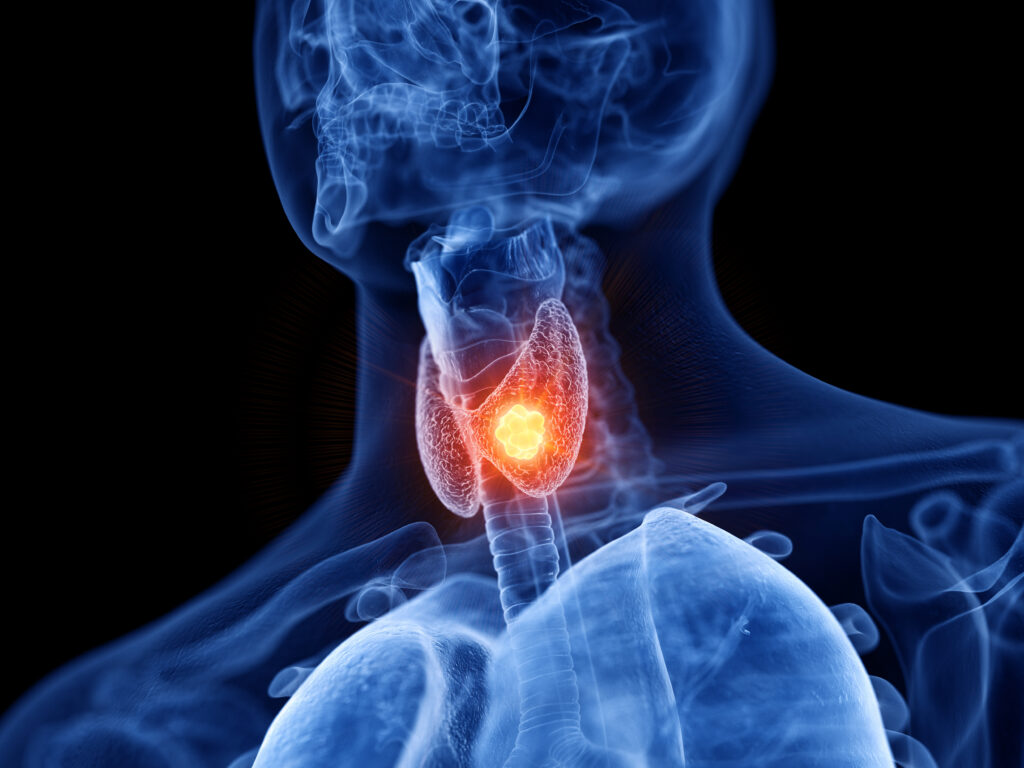
Thyroid cancer presents as a thyroid nodule detected by palpation and more frequently by neck ultrasound. While thyroid nodules are frequent (–50% depending on the diagnostic procedures and the age of the patient), thyroid cancer is rare (only 5% of all thyroid nodules). FNAC should be performed in any thyroid nodule greater than 1cm and in those smaller than 1cm if there is any clinical (history of head and neck irradiation, positive family history of thyroid cancer, suspicious features at palpation, presence of cervical adenopathy) or ultrasonographical suspicion of malignancy (hypoechogenicity, microcalcifications, absence of peripheral halo, irregular borders and regional lymphadenopathy). The results of FNAC are very sensitive for the differential diagnosis of benign and malignant nodules, although there are limitations: inadequate samples and follicular neoplasia. In the event of inadequate samples FNAC should be repeated while in the case of follicular neoplasia, with normal TSH and ‘cold’ appearance at thyroid scan, surgery should be considered.4–7 Considering that the prevalence of indeterminate FNAC is significant (10–25%), several molecules involved in the carcinogenic process have been proposed as markers of thyroid malignancy, but none is recommended because of insufficient data.8,9
Initial Treatment
The initial treatment for DTC is total or near-total thyroidectomy whenever the diagnosis is made before surgery, regardless of nodule size. Less extensive surgical procedures may be accepted in cases of unifocal DTC diagnosed at finally histology after surgery performed for benign thyroid disorders provided that the tumour is small, intrathyroidal and of favourable histological type (classic papillary or follicular variant of papillary or minimally invasive follicular). Surgery is usually followed by the administration of 131I activities aimed at ablating any remnant thyroid tissue and eventually microscopic residual tumour. This procedure decreases the risk of locoregional recurrence and facilitates the long-term surveillance based on serum Tg measurement and diagnostic radioiodine whole body scan (WBS). In addition, the high activity of 131I allows a highly sensitive post-therapeutic WBS to be obtained. Radioiodine ablation is recommended in high- and low-risk patients, while there is no indication in very low-risk patients (those with unifocal T1 tumours less than 1cm in size, with favourable histology, no extrathyroidal extension and lymph node metastases).6 Traditionally, withdrawal of thyroid hormone has been used to optimise the trapping and retention of radioiodine by increasing endogenous TSH levels. The consequent state of hypothyroidism is unpleasant for most patients and sometimes results in marked morbidity and impairment of quality of life.10 Since 1995, rhTSH has been employed in clinical trials for post-surgical thyroid remnant ablation in DTC patients. A study published in 2001 demonstrated 100% ablation using 131I activity determined by individual dosimetry (average dose of 110.3mCi).11 Recently, a randomised, multicentre, controlled study12 designed to investigate whether preparation of patients with rhTSH while on levothyroxine suppressive (LT4) therapy was equivalent to preparation by LT4 withdrawal demonstrated comparable remnant ablation rates (100%) in patients treated with a fixed dose of 100mCi of 131I (3,700 MBq) by either administering rhTSH or withholding thyroid hormone. rhTSH-prepared patients maintained a higher quality of life13 and received less radiation exposure to the body.14 Based on these results, the use of rhTSH for thyroid remnant ablation has been approved by the European Medicines Agency (EMEA) in February 2005 and by US Food and Drug Administration (FDA) in December 2007. More recently, another randomised prospective study employing rhTSH preparation for thyroid ablation found that 50mCi and 100mCi of 131I were equally effective even in the presence of lymph node metastases.15 Based on this evidence, rhTSH may be considered to be the treatment of choice for post surgical thyroid ablation.
Short-term Follow-up
Six to 12 months after initial treatment, follow-up aims to ascertain whether the patient is free of disease, and is based on physical examination, neck ultrasound, serum Tg measurement and diagnostic 131I WBS.6,7 Currently, most of the patients (nearly 80%) will be in complete remission as indicated by no clinical evidence of residual disease, unremarkable neck ultrasound and undetectable basal and stimulated serum Tg (in the absence of serum Tg antibodies).
A diagnostic 131I WBS does not add any clinical information in this setting and may be omitted. In association with a negative post-therapy WBS obtained at the time of thyroid remnant ablation, these findings are sufficient to classify the patient as free of disease, and the rate of subsequent recurrence in this group is very low (<1.0% at 10 years).16,17 These patients may be shifted from suppressive to More recently, another randomised prospective study employing rhTSH preparation for thyroid ablation found that 50mCi and 100mCi of 131I were equally effective even in the presence of lymph node metastases. replacement LT4 therapy, with the goal of maintaining serum TSH level within the normal range. A significant proportion of patients defined as high-risk at the time of diagnosis may appear to be free of disease at their first follow-up after initial treatment. However, the risk of relapse in the long term may be significant; therefore, it is advisable to maintain these patients on suppressive doses of LT4 therapy (TSH around 0.1µUI/ml) for a further three to five years.18 Long-term Follow-up
The subsequent follow-up of patients considered to be free of disease at the time of their first follow-up will consist of physical examination, The subsequent follow-up of patients considered to be free of disease at the time of their first follow-up will consist of physical examination, basal serum Tg measurement on LT4 therapy and neck ultrasound once yearly. basal serum Tg measurement on LT4 therapy and neck ultrasound once yearly. No other biochemical or morphological tests are indicated unless some new suspicion arises during evaluation.
The question of whether a second rhTSH-stimulated Tg test should be performed in disease-free patients is a matter of debate. Recent studies reported that this procedures has little clinical utility in patients who had no biochemical (undetectable serum Tg) or clinical (imaging) evidence of disease at the time of their first rhTSH-Tg. In this group, the second test confirmed complete remission in almost all patients.19,20
The small subgroup of patients with evidence of persistent disease after initial treatment may require further treatment. When the disease is limited to locoregional lymph nodes the option is between additional surgery or 131I treatment (if there is 131I uptake in their metastases). Distant metastases are best treated by surgery whenever possible (resectable isolated localisation). When surgery is not feasible, radioiodine is the treatment of choice if the neoplastic tissue retained the expression of the sodium–iodide symporter (NIS) gene. Evidence has shown that distant metastases particularly in the lung are treated by radioiodine in about 40% of patients.20 After cumulative doses of 22GBq of 131I, further treatment is usually ineffective.21
During the evaluation of metastatic patients, positron emission tomography with 18F-fluorodeoxyglucose (18FDG-PET) scanning is gaining attention as a diagnostic and prognostic tool.22 131I WBS-negative and 18FDG-PET-positive patients indicate a group of patients with more aggressive and less differentiated disease carrying a worse prognosis with respect to 131I-WBS-positive and 18 FDG-PET-negative patients, who have less aggressive disease and better prognosis.
Targeted Therapy
Diffuse metastatic disease refractory to radioiodine may be treated with systemic chemotherapy, but unfortunately the results are usually very poor and disappointing.6 Other currently available therapies are only palliative. For several years novel drugs developed to inhibit the function of specific oncoproteins (targeted therapies) have been intensively studied, aiming at shutting down the uncontrolled growth of neoplastic cells without harming normal cells. Papillary thyroid cancer is characterised by mutations leading to constitutive activation of multiple effectors that signal via the MAP kinase pathway. Molecules that block kinase activity at distal steps in the MAP kinase pathway are logical candidate drugs for papillary thyroid cancer.
Several drugs inhibiting tyrosine kinase (TK) receptors have already been used successfully and some of them have enter clinical practice (i.e. imatinib in chronic myeloid leukaemia). TK inhibitors being tested against DTC in clinical trials include motesanib diphosphate, axitinib, gefitinib, sorafenib and sunitinib. None of these is specific for one oncogene protein, but they all target several TK receptors and pro-angiogenic growth factors. The results of phase II–III clinical trials conducted so far are promising.
Administration of motesanib diphosphate, an agent that targets vascular endothelial growth factor (VEGF), platelet-derived growth factor receptor (PDGFR), KIT and RET receptor tyrosine kinases (RTKs), in a single-arm multicentre phase II trial resulted in partial response (PR) and stable disease (SD) rates of 14 and 67%, respectively, in 93 subjects with DTC and tolerable and manageable toxicities.23 Axitinib, an agent that targets all VEGF receptors, PDGFR-β and KIT, was evaluated in a single-arm study of 60 subjects with advanced thyroid cancer and reported PR and SD rates in 13 patients (22%) and in 30 patients (50%), respectively.24 Gefitinib, a small-molecule inhibitor of the EGFR tyrosine kinase, was used to treat 27 patients with radioiodine-refractory, locally advanced or metastatic thyroid cancer of different histotypes, including papillary, follicular, medullary and Hurthle cell carcinoma. No objective responses were observed, although tumour reduction that did not meet the criteria for partial response was achieved in 32% of the patients and five patients with stable disease had a decrease in thyroglobulin to <90% of baseline. In addition, stabilisation of the disease was obtained in 48, 24 and 12% of the patients after three, six and 12 months of treatment, respectively. The authors interpreted these results as modest but still suggestive of a biological effect of the drug.25
The administration of sorafenib in 30 patients with metastatic, iodine-refractory thyroid carcinoma determined a PR in seven patients (23%), a SD in 16 patients (53%) and showed an overall clinical benefit rate (partial response plus stable disease) of 77%, a median progression-free survival of 79 weeks and an overall acceptable safety profile.26 Sunitinib is a inhibitor of VEGFR-1, VEGFR-2, foetal liver tyrosine kinase receptor 3 (FLT3), KIT and PDGFR in both biochemical and cellular assays, but the response rate in clinical trials is still being evaluated.
Conclusion
The preliminary results of these trials are promising and indicate that targeted therapy might become the first-line treatment of metastatic refractory thyroid cancer patients in the near future.


